Many liver cancer patients who are not eligible for surgery or other treatment options have a choice.

Case Review
Liver Cancer Treatment Case 1:
Patient: Male, primary liver cancer
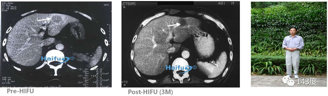
World’s first HIFU treatment for liver cancer, survived for 12 years.
Liver Cancer Treatment Case 2:
Patient: Male, 52 years old, primary liver cancer
After radiofrequency ablation, residual tumor identified (tumor close to the inferior vena cava). Following a second HIFU treatment, complete ablation of the residual tumor achieved, with intact protection of the inferior vena cava.
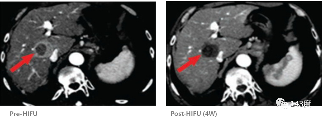
Liver Cancer Treatment Case 3:
Primary liver cancer
Follow-up after two weeks of HIFU treatment showed complete disappearance of the tumor!
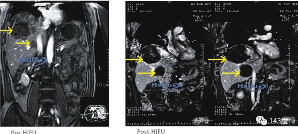
Liver Cancer Treatment Case 4:
Patient: Male, 33 years old, metastatic liver cancer
One lesion found in each lobe of the liver. HIFU treatment performed simultaneously, resulting in tumor necrosis and absorption three months post-surgery.
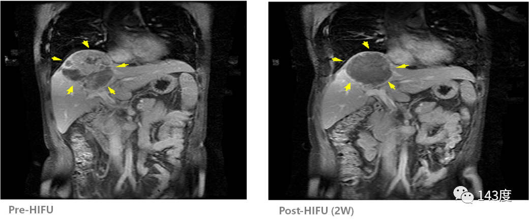
Liver Cancer Treatment Case 5:
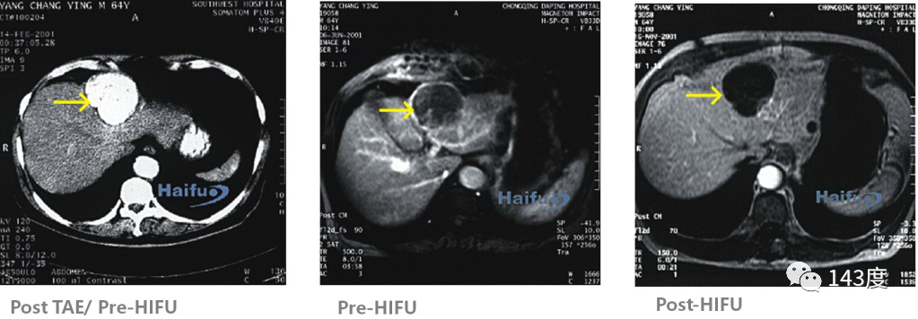
Patient: Male, 70 years old, primary liver cancer
Residual tumor observed on MRI after iodine oil deposition following transarterial embolization. Patchy enhancement disappeared after HIFU treatment, indicating complete tumor ablation.
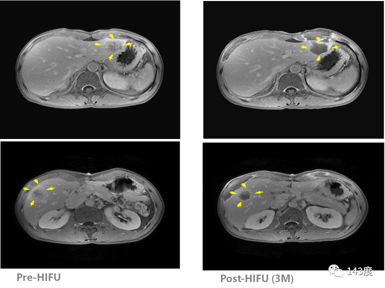
Liver Cancer Treatment Case 6:
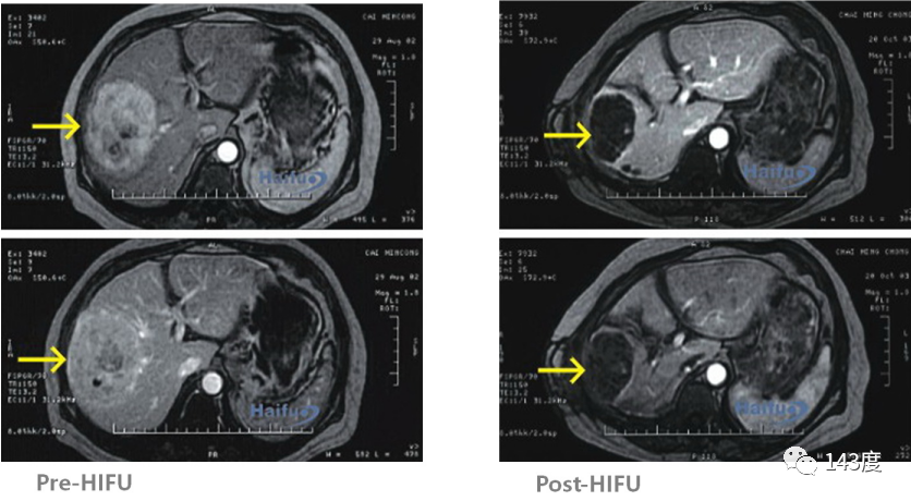
Patient: Female, 70 years old, primary liver cancer
Highly vascular tumor measuring 120mm * 100mm found in the right lobe of the liver. Complete tumor ablation achieved after HIFU treatment, gradually absorbed over time.
Liver Cancer Treatment Case 7:
Patient: Male, 62 years old, primary liver cancer
Lesion located next to the diaphragmatic roof, inferior vena cava, and portal vein system. After 5 sessions of radiofrequency and 2 sessions of TACE, residual tumor identified on follow-up MRI. HIFU treatment successfully inactivated the tumor while preserving the surrounding blood vessels.
Liver Cancer Treatment Case 8:
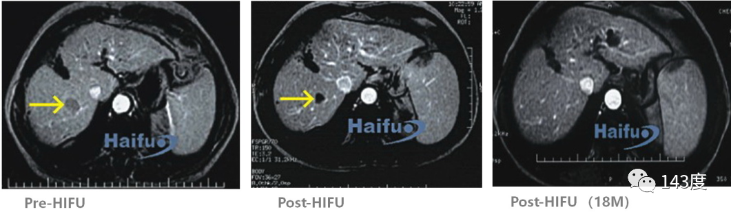
Patient: Male, 58 years old, primary liver cancer
Recurrence observed after surgery for right lobe liver cancer. Complete tumor ablation achieved with HIFU treatment, confirmed by tumor absorption 18 months later.
Hyperthermia for of Liver Cancer – Standardized Research
HIFU (High Intensity Focused Ultrasound) can be used to treat liver cancer. Traditional treatment methods for liver cancer include surgical resection, transarterial embolization, and chemotherapy. However, many patients are diagnosed at an advanced stage or have tumors near major blood vessels, making surgery impractical. Additionally, some patients cannot undergo surgery due to their physical condition, and surgical procedures themselves carry the risk of complications.
HIFU treatment for liver cancer offers several advantages: it is minimally invasive, causes minimal pain and damage, is safe, has fewer complications, and can be repeated if necessary. It can improve patient symptoms and prolong their survival.
Post-HIFU treatment, no cases of tumor rupture, jaundice, bile leakage, or vascular injury have been reported, indicating that the treatment is safe.
(1) Indications: Palliative treatment for advanced tumors, solitary liver cancer on the right lobe with a diameter less than 10cm, huge tumors on the right lobe with satellite nodules that remain confined to the right liver mass, local recurrence after surgery, portal vein tumor thrombus.
(2) Contraindications: Patients with cachexia, diffuse liver cancer, severe liver dysfunction in the late stage, and distant metastasis.
(3) Treatment process: Patients with tumors on the right lobe should lie on their right side, while those with tumors on the left lobe are typically placed in a supine position. Before the procedure, ultrasound is used to locate the tumor for precise targeting and treatment planning. The tumor is then treated through a process of successive ablations, starting from individual points and progressing to lines, areas, and finally the entire tumor volume. Treatment is typically done once a day, with each layer taking approximately 40-60 minutes. The process continues daily, layer by layer, until the entire tumor is ablated. After treatment, the treated area is examined for any skin damage, followed by an external ultrasound scan of the entire target area to assess the treatment efficacy.
(4) Post-treatment care: Patients are monitored for liver function and electrolyte levels. Supportive treatment should be provided for patients with poor liver function, ascites, or jaundice. Most patients have normal body temperature during treatment. A small number of patients may experience a mild increase in temperature within 3-5 days, usually below 38.5℃. Fasting is typically recommended for 4 hours after treatment, while patients with left lobe liver cancer should fast for 6 hours before gradually transitioning to a liquid diet. Some patients may experience mild upper abdominal pain for 3-5 days after treatment, which gradually resolves on its own.
(5) Evaluation of effectiveness: HIFU can destroy liver cancer tissue, causing irreversible necrosis of cancer cells. CT scans show a marked decrease in CT attenuation values within the target areas, and contrast-enhanced CT confirms the absence of arterial and portal venous blood supply to the target area. An enhancement band may be observed at the treatment margin. MRI visualizes changes in signal intensity of the tumor on T1 and T2-weighted images and demonstrates the disappearance of blood supply to the target area in the arterial and portal venous phases, with a delayed phase showing an enhancement band along the treatment margin. Ultrasound monitoring shows a gradual reduction in tumor size, disappearance of blood supply, and tissue necrosis that eventually gets absorbed.
(6) Follow-up: In the first two years after treatment, patients should have follow-up visits every two months. After two years, follow-up visits should occur every six months. After five years, an annual check-up is recommended. Alpha-fetoprotein (AFP) levels can be used as an indicator of tumor recurrence. If the treatment is successful, the tumor will either shrink or disappear completely. In cases where the tumor is still present but no longer contains viable cells, caution should be exercised when a tumor with a diameter greater than 5cm is visible on imaging, and PET scans may be utilized for further clarification.
Clinical observation of pre- and post-treatment results, including alpha-fetoprotein levels, liver function, and MRI scans, have shown a clinical remission rate of over 80% for liver cancer patients treated with HIFU. In cases where blood supply to liver tumors is rich, HIFU treatment can be combined with transarterial intervention. Prior to HIFU treatment, transcatheter arterial chemoembolization (TACE) can be performed to block the blood supply to the central tumor area, with the embolic agent serving as a tumor marker to aid in HIFU targeting. Iodine oil alters the acoustic impedance and absorption coefficient within the tumor, facilitating energy conversion at the HIFU focus and improving.
Post time: Aug-08-2023